What Is an EEG exam (Electroencephalogram )?
An electroencephalogram (EEG) is a non-invasive diagnostic test used to measure and record the electrical activity of the brain. It monitors nerve impulses produced by brain cells (neurons) and helps physicians analyze normal and abnormal brain function for both clinical and research purposes.
EEG is widely used in neurology to evaluate brain disorders, especially epilepsy, sleep disorders, and various neurological conditions.
How Does an EEG Work?
Electroencephalography is a specialized neurophysiological technique that records cerebral electrical activity using electrodes placed on the scalp. In rare and specific cases—such as pre-surgical evaluation for epilepsy—electrodes may be placed directly on or inside the brain.
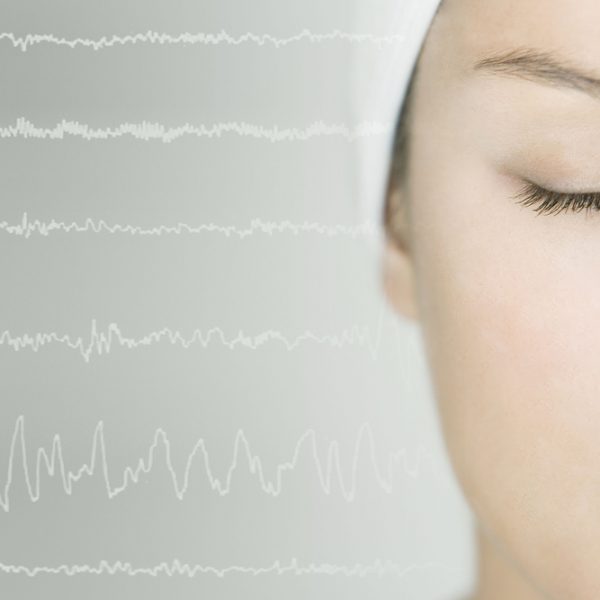
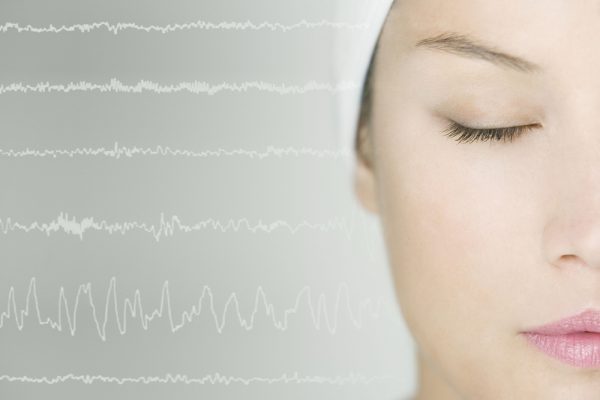
The EEG device detects tiny voltage changes generated by neuronal activity. These signals are amplified, digitized by a computer, and displayed as wave patterns on a screen or printed record, forming the EEG tracing.
Brain Waves Recorded by EEG
An EEG records different types of brain waves, classified by their frequency (measured in hertz, Hz):
- Delta waves (up to 4 Hz) – associated with deep sleep
- Theta waves (4–8 Hz) – common in drowsiness and early sleep stages
- Alpha waves (8–12 Hz) – seen during relaxed, awake states
- Beta waves (above 12 Hz) – linked to active thinking and alertness
Analyzing these brain wave patterns helps clinicians understand brain function and detect abnormalities.
When Is EEG Used?
Electroencephalography is performed under both normal conditions (wakefulness and sleep) and pathological conditions, including:
- Epilepsy and seizure disorders
- Encephalopathy
- Stroke
- Brain injuries
- Structural brain abnormalities
- Altered states of consciousness
EEG is considered a key diagnostic tool for epilepsy, helping confirm the diagnosis and classify seizure types.
Limitations of EEG
Despite its value, EEG has several important limitations:
Spatial Limitations
EEG primarily records electrical activity from the outer layers of the brain (cerebral cortex) and reflects signals from only about one-third of total brain activity.
Time Limitations
Standard EEG recordings are usually limited to 20–30 minutes, which may miss intermittent or rare abnormalities unless extended monitoring is used.
Functional Limitations
Compared to advanced imaging techniques such as PET (Positron Emission Tomography) and MRS (Magnetic Resonance Spectroscopy):
- EEG cannot precisely localize specific brain regions where neurotransmitters or drugs are active
- EEG is less effective in detecting activity in deep brain structures