What is electroencephalogram (EEG)?
Electroencephalogram (EEG) is an monitoring method to record the nerve activity of the brain. Based on the results of these records, further analysis and interpretation of normal and pathological brain activities for research and diagnostic purposes will be done.
Electroencephalography is a special neurophysiological method which registers the cerebral electrical activity through electrodes (probes) placed on the outside of the head (skull) or inside the of the brain (this method is used for preparation in patients with epilepsy).
The diagram of an electroencephalogram (EEG), is obtained as a result of a voltage change and with use of a computer, it converts the signal to digital which is showed on a list of paper.
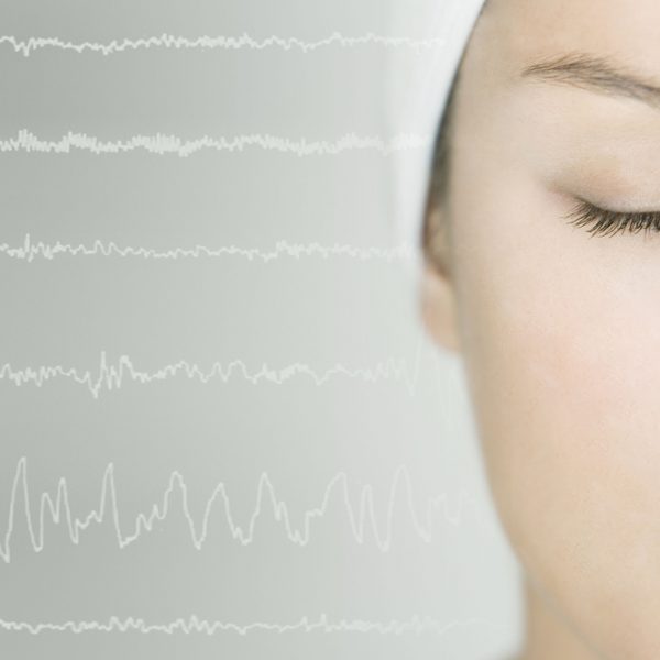
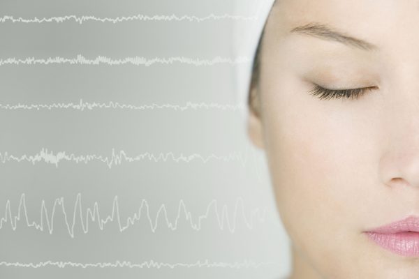
EEG can record the brain waves
EEG records brain waves which are separated according to frequency of:
- delta waves (up to 4 hertz)
- theta wave (4 to 8 Hz)
- alpha waves (8 to 12 Hz)
- beta waves (over 12 Hz)
When is applied?
Electroencephalography is applied in normal conditions (alertness, sleep) and pathological conditions (epilepsy, encephalopathy, stroke or other structural errors) and is used as necessary diagnostic method for testing and diagnosing epilepsy.
Are there any limitations?
Unfortunately, EEG has its significant limitations. They are relate to the spatial (EEG electrical activity can observes on only 1/3 of the whole brain) and time (EEG under standard conditions can be used only for 20 minutes on one subject).
There are also some disadvantages. Unlike the PET and MRS, the electroencephalogram cannot recognize specific locations in the brain at which various neurotransmitters, drugs, etc. can be found. Also, EEG poorly measures the neural activity that take place below the upper layers of the brain (the cortex).


